Right renal mass, a rare disease and diagnostic dilemma in a young female
DOI:
https://doi.org/10.59058/jaimc.v21i2.126Keywords:
Extra skeletal Mesenchymal Chondrosarcoma, Renal Extra skeletal Mesenchymal Chondrosarcoma.Abstract
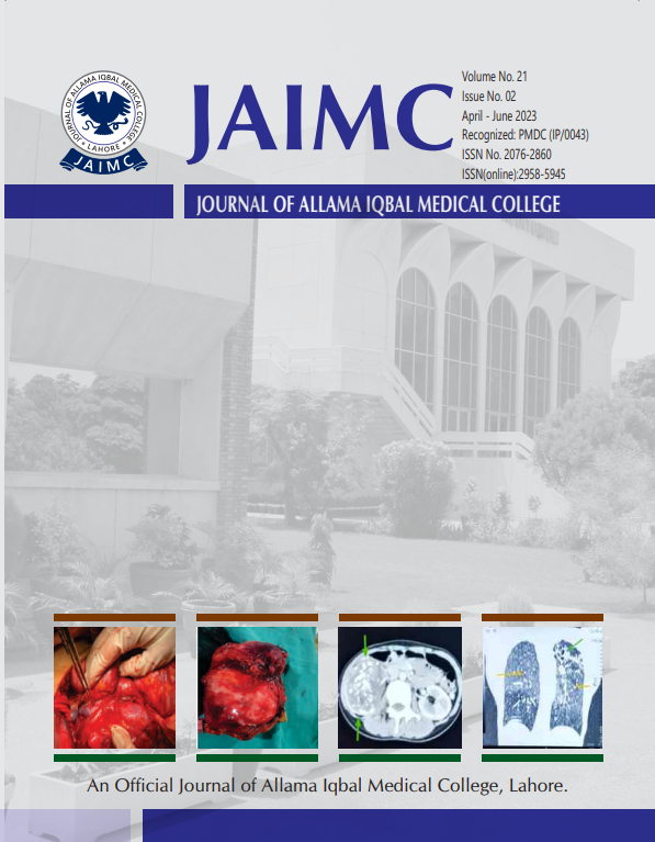
A 22-year-old female from Afghanistan presented with history of pain right lumbar region along with feeling of heaviness and weight loss. The abdominal examination revealed a mass approximately 10*10 cm in the right lumbar region which was bimanually palpable. The diagnosis of Right lumbar mass was made on ultrasound. Final CT scan confirmed its origin from right kidney along with metastasis in lung. The ultrasound guided biopsy turned out to be spindle cell tumour of kidney. Right sided nephrectomy along with removal of proximal ureter done. Histopathology confirmed the diagnosis of mesenchymal chondrosarcoma of right kidney. Postoperative outcome of patient was uneventful.
Downloads
Published
How to Cite
Issue
Section
License
Copyright (c) 2023 Faryal Azhar, Mudassar Niaz, Zakir Sial

This work is licensed under a Creative Commons Attribution 4.0 International License.
The articles published in this journal come under creative commons licence Attribution 4.0 International (CC BY 4.0) which allows to copy and redistribute the material in any medium or format Adapt — remix, transform, and build upon the material for any purpose, even commercially under following terms.
-
Attribution — You must give appropriate credit, provide a link to the license, and indicate if changes were made. You may do so in any reasonable manner, but not in any way that suggests the licensor endorses you or your use.
- No additional restrictions — You may not apply legal terms or technological measures that legally restrict others from doing anything the license permits.
The editorial board of the Journal strives hard for the authenticity and accuracy of the material published in the Journal. However, findings and statements are views of the authors and do not necessarily represent views of the Editorial Board. Many software like (Google Maps, Google Earth, Biorender (free version)) restricts the free distribution of materials prepared using these softwares. Therefore, authors are strongly advised to check the license/copyright information of the software used to prepare maps/images. In case of publication of copyright material, the correction will be published in one of the subsequent issues of the Journal, and the authors will bear the printing cost.


